The 3D geometry is crucial when a clinician grades a
stenosis, measures a bifurcation angle, or steers a
guidewire past a side branch. However, X-ray angiography only provides
sparse 2D projections of the deforming vessels, each from a different
angle at a different moment. In practice, clinicians have to infer the 3D geometry
from these 2D projections, which depends on expertise. Additionally,
the 3D geometry and its mapping to the 2D image are
indispensable to the development of robotic endovascular systems.
The fundamental challenge arises from the temporal variability. More specifically,
cardiorespiratory motion deforms the vessels throughout the acquisition. And X-ray
angiography is performed one view at a time, which means that views acquired at different
times do not correspond to the same 3D geometry, breaking the multi-view consistency. This
variability, combined with the constraints of the imaging protocol, results in a severely
ill-posed inverse problem: reconstructing a slender, temporally evolving 3D geometry from
sparse and temporally misaligned 2D projections. In this problem setting, it is infeasible to
exploit multi-view consistency or to obtain 3D ground truths for training on real-world data,
while a synthetically trained model must carefully handle the sim-to-real gap.
In this context, we propose an automated dynamic cardiovascular reconstruction method called
AutoCAR, which includes the following key designs.
Sparse backward projection. It leverages the sparsity of vascular
structures by treating cardiac vessels as one-dimensional manifolds in 3D space, and
ensures detail preservation by restricting computation and storage to those manifolds.
Pose domain adaptation. It utilizes the viewing preferences of human
experts in XA imaging, and extends the domain adaptation technique from the image domain
to the pose domain to ensure the generalizability of a synthetically trained model in
real-world scenarios.
Vascular graph optimization. It leverages the curve-network
characteristics of vessels for robust vascular topology and temporal coherence.
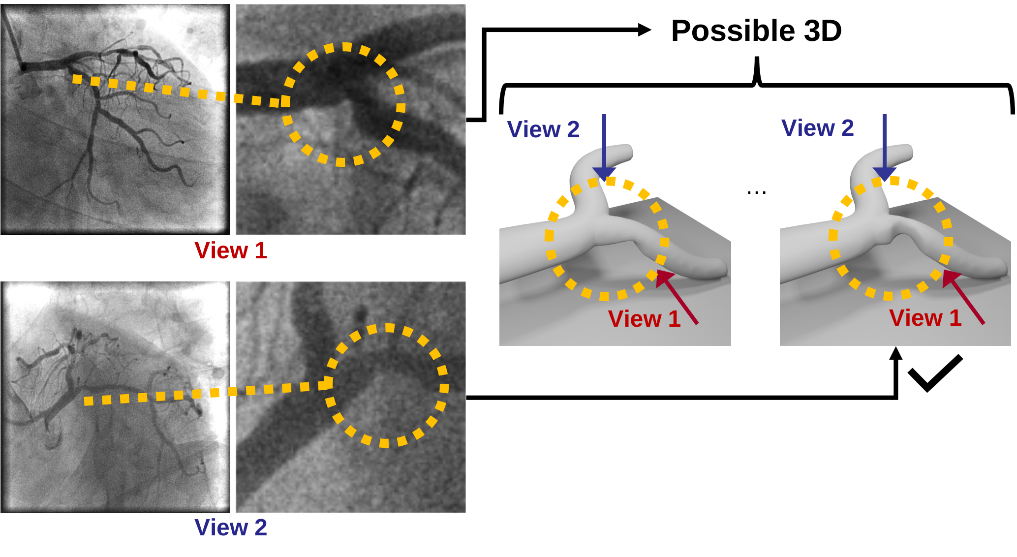
A stenosis in one view is consistent with many 3D shapes, and they disagree about the one
quantity being graded — cross-sectional area. A second view does not hand the answer over
either. Two questions have to be settled by eye first: where is the same site in view 2, and
what 3D shape do the two views agree on?
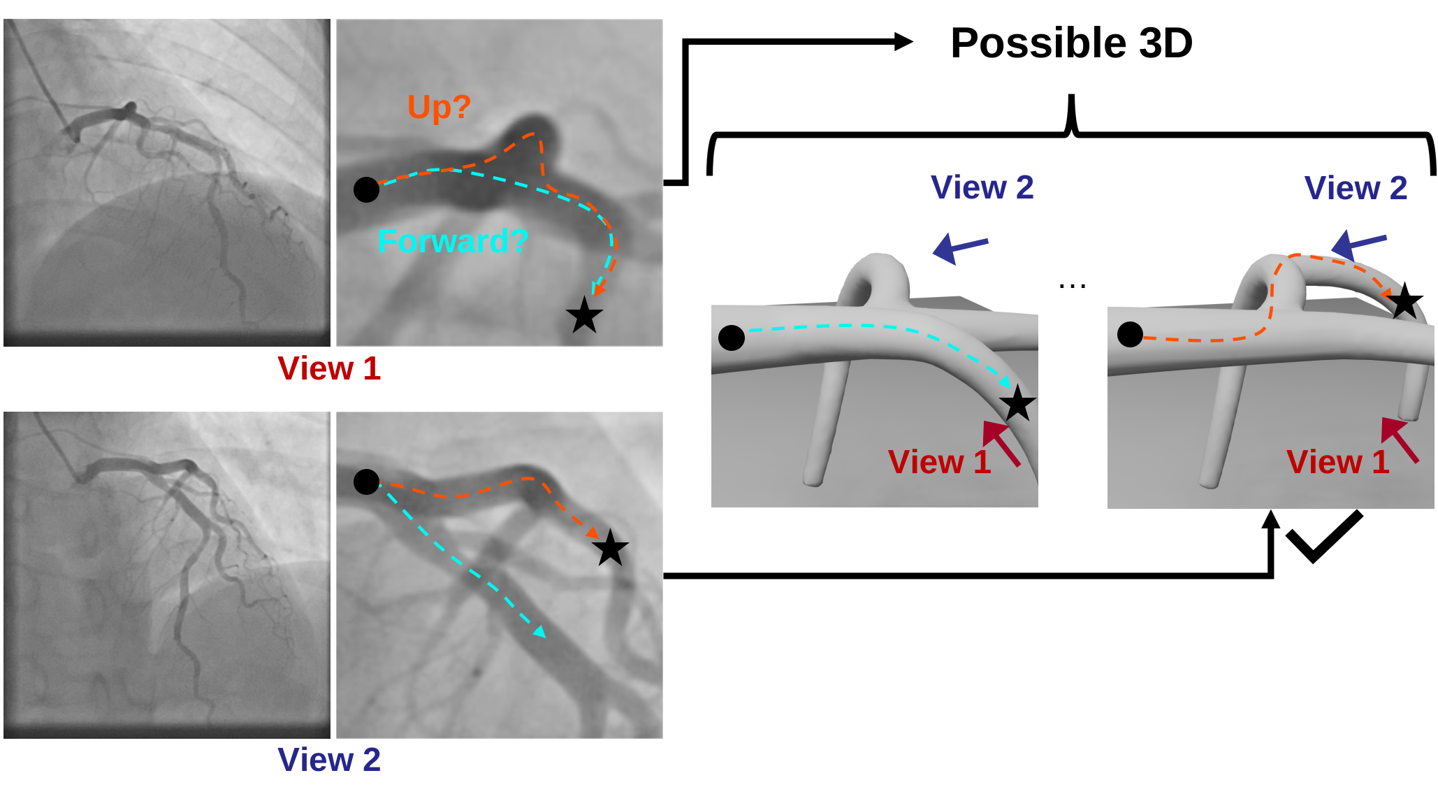
A start and a target in one view leave the 3D topology open: several routes project to the
same picture, and they call for different moves — up rather than forward. Resolving which
one is what the second view is for, and it is the same correspondence problem as above,
asked about topology instead of severity.
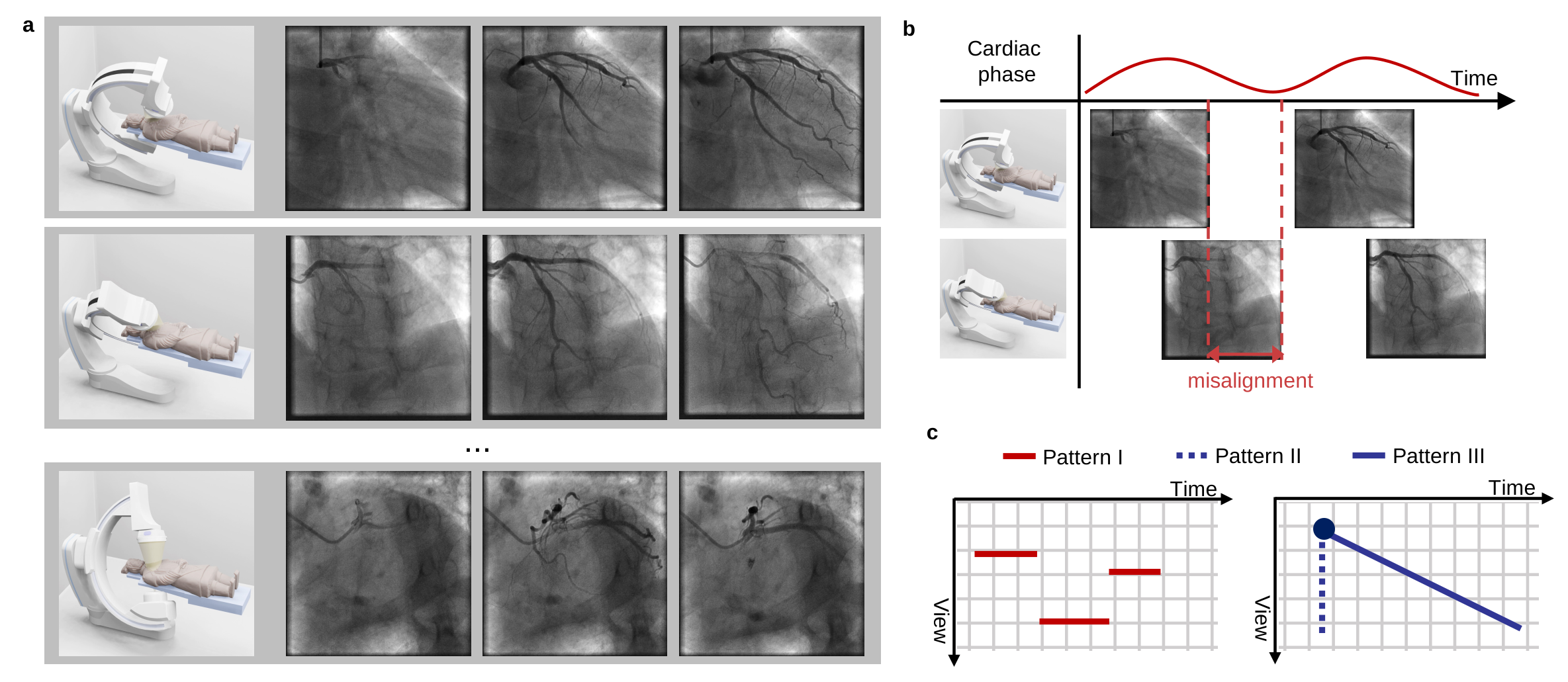
Each row is one viewing pose. The protocol records a video from a fixed viewpoint and only
then moves the C-arm to the next — it is not one continuous sweep, so a frame in one view
and a frame in another are different instants. ECG gating narrows that mismatch without
removing it. Capturing views within a sixth of a cardiac cycle would need the C-arm to
rotate at 1080 deg/s and record at 900 fps, against 60 fps on current systems.
Extended Data Fig. 3.
The reconstruction is what a navigation system consults: the 3D geometry together with its
mapping back into each 2D view, recovered frame by frame.
Demos
Drag inside a 3D panel to orbit it. Move the pointer across a 2D panel to wipe between the
original angiogram and the reprojected reconstruction.
The reconstructed centreline deforms in step with the two X-ray views it was recovered from,
and 2D–2D and 2D–3D correspondences are colour coded.The same colours are baked into the reprojected stills and clips, so a vessel segment keeps
its colour across the 3D view and both 2D panels.
The 3D centreline, instead of surfaces, is shown because of the browser's limitations on
dynamic surface meshes. As a compensation, static surface meshes at a single time step are
shown underneath. Please find
more cases (dynamic centreline),
more cases (static surface), and
comparison with existing works, and
failure cases.
References
Zhu, Y., Wang, Y., Di, C., Liu, H., Liao, F. & Ma, S. Sparse and transferable
three-dimensional dynamic vascular reconstruction for instantaneous diagnosis.
Nature Machine Intelligence7, 730–742 (2025).
doi:10.1038/s42256-025-01025-7
Zhao, H. et al. Self-supervised learning enables 3D digital subtraction angiography
reconstruction from ultra-sparse 2D projection views: a multicenter study.
Cell Reports Medicine (2022).
Banerjee, A., Galassi, F., Zacur, E., De Maria, G. L., Choudhury, R. P. & Grau, V.
Point-cloud method for automated 3D coronary tree reconstruction from multiple
non-simultaneous angiographic projections.
IEEE Transactions on Medical Imaging39, 1278–1290 (2020).
Ying, X., Guo, H., Ma, K., Wu, J., Weng, Z. & Zheng, Y. X2CT-GAN: reconstructing CT
from biplanar X-rays with generative adversarial networks.
Computer Vision and Pattern Recognition (2019).
Wang, L. et al. Weakly-supervised 3D coronary artery reconstruction from two-view
angiographic images. arXiv (2020).
Feldkamp, L. A., Davis, L. C. & Kress, J. W. Practical cone-beam algorithm.
Journal of the Optical Society of America A1, 612 (1984).
doi:10.1364/JOSAA.1.000612
How to cite
@article{zhu2025autocar,
  title   = {Sparse and transferable three-dimensional dynamic vascular
             reconstruction for instantaneous diagnosis},
  author  = {Zhu, Yinheng and Wang, Yong and Di, Chunxia and Liu, Hanghang
             and Liao, Fangzhou and Ma, Shaohua},
  journal = {Nature Machine Intelligence},
  volume  = {7},
  number  = {5},
  pages   = {730--742},
  year    = {2025},
  doi     = {10.1038/s42256-025-01025-7},
}
Acknowledgements
We thank Yanping Yang and Zhiqiang Guo for XA pre-processing advice; Keer Hu for advice on
visualization; Yichao Zheng for clinical advice; and Haohan Xu, Zhengyang Zhang and Linxiao
Yang for discussion. This work was supported by the National Natural Science Foundation of
China (grant numbers 32371470, 82341019, S.M.), the National Key Research and Development
Program of China (2024YFA0919800, S.M.), the Department of Science and Technology of
Guangdong Province (grant number 2023B0909020003, S.M.), and the Cross-disciplinary
Research and Innovation Fund of Tsinghua SIGS (grant number JC2022007, S.M.).


 NMI 7(5), May 2025.
 Extended Data Fig. 3